In this post we discuss a topic
that everyone would have heard before. Let's talk about migraines, a disorder
that involves a huge physiological and molecular background, and it has an
alarmingly high impact worldwide. We are going to talk about the etiology of
the disease, the molecular and supramolecular processes that trigger it and we
will end talking about traditional therapies, the most novel therapies and, of
course, prophylaxis or prevention of migraines. This post is not intended to be
a review, but a somewhat more scientific explanation that what is generally
known about migraines, explained so that everyone can understand and enjoy its
content.
What are migraines?
Migraine is a common and incapacitating
neurovascular disorder, primarily genetic in origin and characterized by very
severe headaches, dysfunction of autonomous nervous system and, in some
patients, the appearance of an aura that encompasses visual, sensory and/or
speech symptoms temporarily. The headache is usually throbbing and appears on
one side of the head, i.e. it is unilateral. According to the World Health
Organization (WHO), migraine has an average prevalence of 10-15% of the
global population. Furthermore, the risk for this disorder is up to three
times higher for women due to certain hormonal influences that we will explain
later. In turn, WHO has classified chronic migraine as one of the most
disabling disorders along with quadriplegia, psychosis and dementia.
Although migraine attacks can start at any
age, the incidence is concentrated mainly in adolescence. The average frequency
is 1.5 attacks per month and the average duration of attacks is about 24 hours
but it can reach 2-3 days if no appropriate treatment is available. In migraine
without aura, also called common migraine, attacks are associated with nausea,
vomiting and excessive sensitivity to light (photophobia), sound or movement. About
65% of patients have common migraine, 20% migraine with aura and 15% both. Some
subjects gradually evolve from episodic migraine to chronic, which
affects 1-2% of the general population and is characterized by 15 days
or more of headache per month. As in the case of epileptic seizures, one of the
more consolidates triggers of migraines is stress. It is important to note that
aura can appear in patients with either episodic or chronic migraines, although
the incidence of aura is higher in the latter. Therefore, chronic is not
synonymous to aura, but to more than 15 days per month of headache. The
following image shows the representation of unilateral headache that is
experienced during a migraine, including visual and auditory area.
www.atlantatmjfacepain.com
Migraines have an important psychological,
social and economic impact. About 75% of patients are functionally impaired
during an attack and more than half of them require assistance from third
parties.The annual economic impact of migraine is estimated at 27 billion euros
in work days lost around Europe.
The management of migraines includes an
adequate pain relief and the reduction or complete elimination of the attacks. Nowadays,
there are several acute and prophylactic treatments for migraines, but a small
proportion of patients remain untreatable and no new treatment or drug has
emerged in recent years to try to solve this problem.
How are migraines originated?
Migraine is a complex condition whose
pathophysiology is not completely understood. The most widely accepted theory
for the initiation and perpetuation of migraine is a combination of vascular
and neural mechanisms. This theory suggests that the headache depends on
the activation of trigeminovascular pain pathway and in patients with
aura, this aura is represented by a wave of neuronal hyperactivity followed by
occipital cortical depression (back of the brain), called cortical spreading
depression or CSD.
Like most solid organs,
the brain is insensitive to pain. The sensitive intracranial structures,
including nociceptors (pain receptors) are on the walls of arteries, veins,
venous sinus and meninges (connective tissue membranes covering the brain). Peripheral
vessels and meninges have sympathetic and parasympathetic sensory innervation. The trigeminovascular
system consists of large intracranial blood vessels innervated by the
ophthalmic branch of the trigeminal nerve. This nerve, also known as the fifth
cranial nerve, has sensory and motor branches, and it is the largest of all
cranial nerves. Among these sensory branches we find the facial and scalp
nociceptors, including the meninges. In the next picture we can see an example
of trigeminovascular system innervation of the meningeal vessels, as well as
the ascension of the sensory pathways to the brain.
www.rayur.com
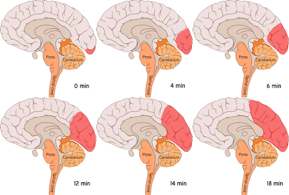
As mentioned above, about 20% of patients with migraine have a neurological
phenomenon called aura. This event has been linked to cortical spreading
depression (CSD), which refers to a wave of neuronal hyperactivity followed by
a long lasting decrease in neuronal activity. Generally, it is
triggeredin the occipital part of the cortex and from there it is slowly
propagated at a speed of 2-5 mm/min towards adjacent tissues. In the following
animation we can see a representation of the wave spreading through the brain.
Creative Commons
Attribution: PJ Lynch, Jaffe CC.
This wave is initiated by a massive increases inextracellular potassium
and the excitatory neurotransmitter glutamate, which can trigger
depolarization or activation of nociceptive neuronal endings of the
trigeminovascular system. Potassium stimulates nerve endings directly, while
glutamate exerts its effect through its neurotransmitter functions. The
increase of potassium and glutamate is explained in part by mutations in
certain ion channels (below). Activation of these nociceptive terminals causes
neuronal depolarization and the subsequent release of vasoactive
neuropeptides such as: CGRP (calcitonin gene related peptide), substance
P and neurokinin A, all of them with both vasodilatory and neuronal
excitation capacity. CGRP can increase the sensitivity of perivascular
nociceptors and vasodilate cranial vessels by stimulating the release of nitric
oxide, a potent vasodilator. Substance P is a mediator that, on one hand
increases the sensitivity of pain-related neuronal endings indirectly by
stimulating blood mast cells to produce histamine (inflammatory mediator) and
on the other hand vasodilates vessels. All these phenomena create a feedback
loop that progressively increases pain. Another neuroanl messenger that plays a
key role in the pathophysiology of migraines is the polypeptide PACAP (pituitary
adenilate cyclase-activating polypeptide), expressed in trigeminal neurons,
whose induction, shown in experimental studies in humans, causes an increase in
extracerebral vessels vasodilation.
www.realclearscience.com
The wave of neuronal
hyperactivity and vasodilation results in a wave of hiperemia
(increasing of blood volume in the brain). This phenomenon is followed by a
wave of oligohemia (decreasing of total volume of blood) that runs
through the cerebral cortex associated with a wave of neuronal depression. The
mechanisms leading to this depression in cortical activity are not entirely
clear but a possible explanation is the decrease of occipital cortical metabolism
by decreased blood volume. This would explain symptoms such as flashing lights
that certain patients experiment in one eye during a migraine attack. Increased
blood volume stimulates the retina, causing the appearance of phosphenes or
bright spots, while the subsequent decrease in blood volume stops this process.
This process is constantly repeated. Despite what it may seem,CSD does not
cause tissue damage in healthy brains.
www.margelee.com
Certain genetic susceptibility or familial
predisposition is involved in the etiology of the disease. We mentioned earlier
that increase in both extracelular potassium and glutamate may be due to mutations
in certain ion channels. There are many genes whose mutation triggers changes
in ion channels leading to the development of the pathophysiology of migraines,
but we will not dwell on many details. As an example, we will talk about Familiar
Hemiplegic Migraine (FHM), an autosomal dominant migraine with an
especially pronounced aura, in which mutations have been found in three
causative genes encoding for ion channels.
- CACNA1A: encodes the α1A subunit of neuronal calcium channel P/Q type. Mutations in this gene result in gain of function of calcium channel Ca 2.1 and the subsequent release of dependent neurotransmitters, including glutamate, from cortical neurons, facilitating the induction and propagation of the CSD and the activation of the trigeminovascular system.
- ATP1A2: Encodes the α2 subunit of ATPase Na+/ K+, expressed in glial cells and involved in extracelular K+ uptake and the production of a Na+ gradient that is used in glutamate recaptation. Mutations in this channel can lead to increased extracellular potassium and glutamate, reducing the CDS threshold.
- SCN1A: encodes the α1 subunit of the voltaje-dependent sodium channel Nav1.1. This channel is critical for the generation and propagation of action potentials. A mutation in this gene may lead to increased excitability of dendrites and neuronal firing.
Therefore, roughly speakin in patients
with common migraine without aura, the headache is produced almost entirely by
activation of trigeminovascular system, and in patients with migraine with aura
both phenomena occur, activation of the trigeminovascular system and cortical
spreading depression (CSD), which positively regulate each other. These events
are produced mainly by genetic factors.
One reason for the fact that the risk of
migraines is 3 times higher in women than in men is the existence of genetic
polymorphisms in the estrogen receptor ESR1, which increase its
activity. This receptor is a transcription factor that is expressed in several
areas of the brain and regulates, among other functions, gene expression
affecting the synthesis of CGRP, serotonin and glutamate. Exacerbated
activation of this receptor stimulates the release of nitric oxide via CGRP
and, in turn, the production of more CGRPs by the action of glutamate.
How can we treat migraines?
Immediate treatment of an attack looks for
a quick elimination of pain. In many patients the administration of analgesics
is sufficient to control pain during an attack, however, some individuals have
a reduced response to pain medication so serotonin receptor 5-HT1B/1D
agonists can be used. Another strategy
to suppress migraine is blocking the release of neuropeptides or the activation
of its receptors.
Analgesics: First treatment
option. These include, aspirin, acetaminophen, ibuprofen, naproxen or
diclofenac.
Agonists of the 5-HT1B/1D receptor: Also called triptans
they should only we considered when there is an inadequate response to
analgesics because they can produce certain side effects. Examples include
eletriptan, sumatriptan, zolmitriptan, naratriptan or rizatriptan. They are
very potent and effective, and exert their action at three levels:
- Cranial vasoconstriction by inhibiting the release of vasodilators due to activation of the serotonin 1B receptor.
- Activation of serotonin 1D receptor, decreasing peripheral pain transmission by inhibiting both peripheral neuronal transmission and trigeminocervical complex activation.
CGRP receptor antagonists: Currently, monoclonal
antibodies against CGRP or its receptor have showed very promising results
in animal models. Nowadays, certain clinical trials are performing
investigations in human patients with monoclonal antibodies and the preliminary
data is very encouraging. However, there are still no definitive conclusions on
such studies.
The prophylaptic options recommended are:
- Beta blockers: antagonists of adrenaline and noradrenaline at presynaptic level. They also inhibit the nitric oxide’s activity.
- Calcium antagonists: they block calcium channels, preventing the release of neurotransmitters such as glutamate.
- Antidepressants; such as amitriptyline, they modulate pain. Prevent the recaptation of serotonin and norepinephrine, increasing their levels in the brain.
- Antiepileptics: such as topiramate, they act on processes that ultimately suppress the initiation and propagation of the CSD.
For all the above,
migraines are a major public health problem that affects a large part of the
population and which is being actively investigated with the aim of discovering
more innovative and effective therapies, as well as to find a solution to
intractable patients. There are still many loose ends at the mechanistic level
in the pathophysiology of migraines, as well as many links between discoveries
and isolated observations, but gradually we will finally understand the complex
neural and vascular network.
Finally we include a
very interesting and informative video created by Novartis that advertize one of
its flagship drugs, Excedrin, very popular to treat migraine attacks. It is
composed of a mixture of aspirin, acetaminophen and caffeine. In this video,
titled “What does a migraine feel like?”, they use a helmet and virtual glasses
specially developed to simulate the symptoms of a migraine attack on healthy
people. Surely you will be surprised. We hope you like it.
For any questions or suggestions do not
hesitate to contact us.
Acknowledgement: Thanks to José
Manuel González-Navajas and Beatriz Lozano-Ruiz from CIBERehd Alicante for
their great support and help in the translation.
REFERENCES:
- World Health Organization (WHO): https://www.who.int/es/
- Brain stem activation in spontaneous migraine attacks. Weiller C, May A, Limmroth V, Jüptner M, Kaube H, Schayck RV, Coenen HH, Diener HC. Nat Med 1995, 1:658-660.
- Migraine-Current understanding and treatment. Goadsby PJ, Lipton RB, Ferrari MD. N Engl J Med 2002, 346(4):257-70.
- Brain Metabolism in migraines. Montagna P, Welch KA. In Olesen J, Goadsby PJ, Ramadan NM, Tfelt-Hansen P, Welch KWA eds. The Headaches 3rd Ed. Philadelphia: Lippincott-Williams and Wilkins. 2006; Chapter 38:363-367.
- Synthesis of migraine mechanisms. Ollesen J, Goadsby PJ. In Olesen J, Goadsby PJ, Ramadan NM, Tfelt-Hansen P, Welch KWA eds. The Headaches, 3rd edition, Philadelphia: Lippincott-Williams and Wilkins. 2006; Chapter 42:393-398.
- Migraine in the triptan era: lessons from epidemiology, pathophysiology and clinical science. Bigal ME, Ferrari M, Solberstein SD, Lipton RB, Goadsby PJ. Headache 2009, Suppl 1:S21-33.
- Changes in functional vasomotor reactivity in migraine with aura. Wole ME, Jäger T, Bäzner H, Hennerici M. Cephalalgia 2009, 29:1156-1164.
- Migraine, neuropathic pain and nociceptive pain: Towards a unifying concept. Chakravarty A, Sen A. Medical Hypothesis. 2010, 74:225-231.
- Activation of central trigeminovascular neurons by cortical spreading depression. Zhang X, Levy D, Kainz V, Noseda R, Jakubowski M, Burstein R. Ann Neurol 2011. 69:855-865.
- Chemical mediators of migraine: Preclinical and clinical observations. Gupta S, Nahas SJ, Peterlin L. Headache 2011, 51(6):1029-45.
- Update on the genetics of migraine. Merikengas KR. Headache Currents 2012; 52: 521-522.
- Evidence-based treatments for adults with migraine. Gooriah R, Nimeri R, Ahmed F. Pain Res Treat 2015, 2015:629382.
- New players in the preventive treatment of migraine. Mitsikostas DD, Rapoport AM. BMC Med 2015, 13:279.
- Migraine in the era of precision medicine. Zhang LM, Dong Z, Yu SY. Ann Transl Med 2016, 4(6):105.


No comments:
Post a Comment